- No products in the cart.
In industries where scientific imaging is used, including biotechnology, medical research and development, and semiconductor industry, experts typically rely on optical and scanning electron microscopes.
Despite performing nearly the same function, there is a major difference between an optical microscope and an electron microscope.
Optical Microscopes for Scientific Imaging
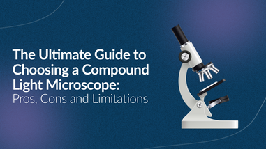
Dating back to the 18th century, optical microscopy involves using one or a series of lenses. The use of optical microscopes characterizes traditional microscopy in that it offers a closer view of a sample through a magnifying lens with visible light.
An optical microscope may either have a converging or a concave mirror. While a converging lens is a commonly used optical instrument, the concave mirror is used to illuminate the sample.
Optical Operation
To understand how an optical microscope forms an image on the lens, it is necessary to understand the concept of focal point and focal length.
-
Focal Point - The focal point is the point of heat when you magnify light onto the ground.
-
Focal Length - Focal length is the distance between the lens and the focal point.
In optical microscopy, the smaller the radius of curvature of the converging lens is, the shorter the focal point will be. This explains why a lens with a large diameter may prove more effective than one with a smaller diameter.

Scanning Electron Microscopes (SEM) for Scientific Imaging
In Scanning Electron Microscopy (SEM), a beam of electron is used to illuminate the sample instead of visible light. The energy-packed electrons when fall on the sample, they emit signals revealing the chemical composition, texture, crystalline structure, and material orientation of the solid sample.
This allows the viewers to notice the effects of electrons as they fall onto the sample.
The Detectors
SEM features the following three types of detectors:
1.Secondary Electron Detector (SED)
When secondary electrons interact with the sample, they create a huge reflection angle. The SED, therefore, is helpful in getting detailed topographical information.
2.Back-Scattered Electron Detector (BSED)
The back-scattered electrons boast a smaller reflection angle, penetrating further into the sample. Hence, the BSED provides the basic topographical and compositional information to the viewers.
3.Energy Dispersive Spectrum Detector (EDS)
The EDS provides the viewers with detailed chemical composition information that neither SED nor BSED can provide.
Kinetic Energy and Signal Emission
The high-power electrons used in SEM produce significant amounts of kinetic energy. As soon as they fall on a solid sample, they emit a number of signals. This electron-sample interaction includes secondary electrons and back-scattered electrons that produce the image, diffracted back-scattered electrons that identify the orientation of crystalline structures, photons used for the analysis of elements and continuum x-rays, heat, and visible light.
Difference between Optical Microscope and Electron Microscope
It's clear these two machines have differences in composition and function, but how does that affect your experiments? When should you use an optical microscope vs. an SEM?
Difficulty Level of Operation
Optical microscopes being the oldest and the simplest of all are super easy to use. With this microscope, you can analyze a sample in water or air and a sample in natural coloring. This makes optical microscopy a less complicated alternative for scientific imaging.
The Scanning Electron Microscope is typically larger and more complicated than an optical microscope but recent developments have bridged this gap too by introducing easy-to-use imaging tool. The high-resolution electron microscopes now allow viewers to get a view of the whole sample, navigating around the sample with just a click.
The Resolution Power
In the study of microscopy, the resolution power of a microscope is the smallest detail that can be seen with it. Typically, the wavelength of the beam directly influences the resolution power of a microscope.
The Scanning Electron Microscopes tend to have higher resolution power than optical microscopes, meaning that they offer a much more detailed view of the solid sample.
Wavelengths
In optical microscopy, the resolution power is limited to visible light and so, the wavelength tends to be 400-700 nanometers. This means that these microscopes can offer a 1500 x magnification only and a minimum resolution of 200 nm.
On the contrary, the beam used in Scanning Electron Microscopes contains electrons with energies thousand times greater than that of visible light. This is why the depth of focus and the resolution power of these advanced microscopes are so higher.
Which Microscopy is better for Scientific Imaging?
Both optical and electron microscopes have their pros and cons. While optical microscopy offers ease of use, the highly focus resolution power of SEM draw experts the other way.
For scientific imaging in industries however, the advanced series of an electron microscope, the Desktop Phenom SEM wins the battle. These microscopes are not only affordable and easy to use but they also offer high-resolution scientific imaging and several other advanced features. Hence, they may be the best microscope for scientific imaging.
For over 40 years, Lab Pro has been committed to delivering a complete laboratory solution by offering the highest quality microscopes and imaging equipment for our customers worldwide. Come visit the biggest Lab Supply showroom in the Bay Area, or contact us online or at 888-452-2776 to learn more.


