- No products in the cart.


As quality control issues in biomedical device manufacturing go, maintaining medical-grade purity of nitinol wires and therapeutic devices is especially difficult. Because nitinol is used in many implantable devices like cardiac stents, the standards for the metal’s quality are extremely high—but meeting that standard can be exceedingly tough.
Nitinol’s shape-shifting properties mean that problems lurking within the metal can be hidden until immediately before a surgeon needs to shape the device to the anatomy of the patient. Because of this, maintaining the quality of nitinol-based implantable devices is a top priority for manufacturers, not to mention a major cost.
Visibly flawed nitinol elements are indications that something has gone dramatically wrong in the manufacturing process, yet identifying small inclusions indicative of more subtle problems can be much more troublesome to locate without the use of advanced microscopy techniques, which can be difficult to scale.
The good news is that for groups who are trying to streamline their nitinol wire or device inspection process, a tool as simple as a common light microscope might be the right solution to quality assurance. Using a light microscope instead of a more complicated QA technique might even save time and money when hunting for the type of inclusions which are most likely to harm a patient being treated with a nitinol device.
What Are Nitinol Surface Inclusions, And Why Do They Matter?
Non-metallic or organic impurities in the composition of nitinol have a high probability of forming a non-metallic inclusion (NIM), especially when the nitinol is configured in a wire, such as in many biomedical implant applications. NIMs lead to more rapid fatigue of the nitinol, which may lead to cracking or fractures if the nitinol undergoes a high enough number of conformational changes. For nitinol applications in implants or stents, identifying NIMs is a critical activity within the quality assurance process because an undetected NIM could easily lead to an implant failure when the patient needs it to be perfectly reliable.
NIMs are not always caused by a problem with manufacturing, however. Inclusions can also occur as a result of corrosion caused by outside forces. While corrosion-induced NIMs are less common than manufacturing impurity-induced NIMs, identifying either type of NIM requires the use of a technique which is well-disposed to the task. Laboratory workers and device manufacturers detect NIMs in several ways:
• Chemical submersion tests
• Electron-scanning microscopy
• Photon-scanning microscopy
• Canonical light microscopy
Of these methods, many manufacturers prefer light microscopy, as it is affordable enough, fast enough, and gentle enough on the nitinol itself to be used for industrial-scale quality assurance in implantable medical devices.
The main argument against using light microscopy is that it is incapable of detecting subsurface inclusions in the nitinol. However, it’s possible that tiny subsurface inclusions do not end up harming patients anyway. In fact, research dating from 2010 found that roughly 80% of implantable nitinol stent failures were caused by fully visible inclusions which were larger than 200 microns in size—well within the range of detection using conventional light microscopy, but far too large to be worth spending the time and money to use a photon or electron-based advanced microscopy technique. This means that detecting the nitinol surface inclusions which are the most hazardous to potential patients downstream will require using the right light microscope for the job.

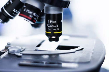
What To Look For In A Microscope For Inspection of Nitinol Devices Or Wires
There are a plethora of different microscopes on the market, but most microscopes are ill-suited for examining nitinol surface inclusions. When the correct microscope is used, it’s easy to see the transition of nitinol in real-time with an abundance of clarity. With the wrong microscope, objective, or stage, however, the chances of robust observation of nitinol’s phase changes are heavily minimized.
The most important features of a microscope intended for inspection of nitinol for the detection of surface inclusions are:
-
Binocular vision
-
A non-reflective stage
-
Top-down lighting
Binocular vision is a staple for all modern microscopes because of the added field of view and ease of focusing which binocular vision allows. In contrast, the location of the microscope’s illumination element is a design choice which helps tailor a microscope to its intended use. Microscopes with the illumination element underneath the stage are excellent for applications which require high magnification of semi-transparent objects, like cells or other biological materials. Because the light must pass through the object being investigated to reach the objective of the microscope, illuminating from below guarantees that no light reflects off of the surface of the sample itself, which is especially important for liquid samples.
Illuminating from the top-down is less common because microscope stages tend to be somewhat reflective, even when they are designed to be non-reflecting. However, illuminating from the top down is also more effective for examining how the object in question reflects light. In the context of nitinol, variation in the reflectiveness of a certain portion of the device or wire would be indicative of a serious inclusion. Likewise, by lighting from above, it is also possible to see shadows cast by imperfections caused by inclusions when the surface of the device is facing the microscope’s objective. With a bottom-up illuminating element, it wouldn’t be possible to detect shadows as easily because the majority of the light would be passing through into the user’s eyes via the unobstructed view of the objective.
This means that the best microscopes for detecting nitinol inclusions are usually not biological microscopes, which utilize bottom-up light sources and a polished stage which is typically at least slightly reflective. Instead, microscopes which are intended for use in industry or in chemistry are better suited to inspecting nitinol because they typically feature top-down lighting and removable or matte stages.
Identifying Imperfections and Inclusions in Nitinol Is Easy With The Right Tools
In conclusion, using industry or chemistry-focused light microscopy is an inexpensive, easy, and effective way to find inclusions in nitinol devices which might be harmful for patients. Rather than spending money on training and equipment for electron or photon microscopy, investigate what kinds of light microscopes your local laboratory supply company can offer your laboratory or manufacturing group. While not all light microscopes will do, the right light microscopes will streamline your nitinol inspection process—and it just might save a patient or two down the road as well.
For over 40 years, Lab Pro has been committed to providing the right microscopes for nitinol inspection work to laboratories in California and worldwide. Come visit the biggest Lab Supply showroom in California, or contact us online or at 888-452-2776 to inquire about our “Be A Lab Pro” inventory management program.